
Anti-mRaspberry Polyclonal IgG Antibody: High-Sensitivity Detection of mFruit-Family Red Fluorescent Proteins
Background on mRaspberry and red fluorescent proteins
mRaspberry is a monomeric red fluorescent protein (RFP) engineered from DsRed (Discosoma sp.) as part of the mFruit series (mCherry, mStrawberry, mPlum, etc.). These derivatives were developed to provide a broad spectral palette of monomeric fluorescent proteins optimized for live-cell imaging, protein tagging, and multicolor microscopy.
The classical mFruit engineering work and FP selection principles are summarized in Shaner et al., “Choosing fluorescent proteins,” from the Tsien lab at the University of California San Diego (UCSD PDF) and in a detailed fluorescent protein user guide from Albert Einstein College of Medicine (Einstein FP guide). tsienlab.ucsd.edu+1
Spectral properties of mRaspberry and related RFPs (excitation ~598 nm, emission ~625 nm) are tabulated in overviews of red and far-red fluorescent proteins from Zeiss / Wake Forest University (Fluorescent proteins – Zeiss campus PDF) and in Wiedenmann et al.’s review on fluorescent proteins for live-cell imaging at Texas A&M University (chem.tamu.edu FP review). microscopy.wfu.edu+1
A more specialized analysis of far-red variants, including mRaspberry, mPlum and E2-Crimson, is available from NCBI PMC and the University of Chicago (e.g. Lin & Tsien, “Autofluorescent proteins with excitation in the optical window,” NIH/NCBI PDF; Strack et al., “E2-Crimson,” UChicago PDF). tsienlab.ucsd.edu+1
These resources frame mRaspberry as a far-red, monomeric DsRed derivative with good compatibility for mammalian expression, deep-tissue imaging and multiplexing.
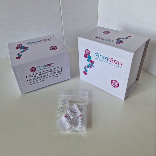
Concept and format of Anti-mRaspberry Polyclonal IgG Antibody
Polyclonal IgG concept
An Anti-mRaspberry Polyclonal IgG Antibody is generated by immunizing a host species (commonly rabbit, goat or sheep) with purified recombinant mRaspberry protein or a fragment containing the structured β-barrel. The immune system produces a diverse polyclonal repertoire of IgG molecules recognizing multiple epitopes across the mRaspberry surface.
General overviews of polyclonal antibody production are provided by:
-
UCLA Animal Research Committee – “Polyclonal Antibody Production” (ucla.edu ARC page) rsawa.research.ucla.edu
-
UC Davis rabbit polyclonal program (ucdavis.edu PDF) heyerlab.ucdavis.edu
-
University of Maryland “Polyclonal Antibody Production Guidelines” (umd.edu PDF) Division of Research
-
UC Berkeley ACUC guidelines for antibodies in laboratory animals (berkeley.edu PDF) acuc.berkeley.edu
These documents describe antigen preparation, adjuvant selection, animal handling and bleed schedules used for high-titer, high-affinity polyclonal IgG production.
Why antibody detection for fluorescent proteins?
Although mRaspberry is intrinsically fluorescent, there are important reasons to detect it with an antibody:
-
Signal amplification – Indirect immunofluorescence with a primary anti-mRaspberry antibody and fluorophore-conjugated secondary antibody can increase the effective signal, which is essential when expression levels are low or when fixation quenches the chromophore. Practical IF amplification guidance is available from Duke University Microscopy Core (IF sample-prep guide) and the University of Nebraska–Lincoln (UNL IF protocol). microscopy.duke.edu+1
-
Harsh fixation / antigen retrieval – Cross-linking fixatives and paraffin embedding can destroy FP fluorescence, but epitopes remain recognizable by antibody. Step-by-step paraffin IF protocols from NCBI / Max Planck and UAMS illustrate how antigen retrieval and permeabilization are combined with antibody detection (IF paraffin protocol, PMC; UAMS immunofluorescence immunotyping). PMC+1
-
Multi-color multiplexing – Anti-mRaspberry can be combined with anti-GFP, anti-mCherry and anti-epitope-tag antibodies to map multiple tagged proteins simultaneously. Multiplex IF strategies are described in protocols from Northwestern University (Bevan lab IF protocol) and Johns Hopkins School of Medicine (multiplex IF chapter). labs.feinberg.northwestern.edu+1
Recent reviews from NCBI PMC highlight that antibodies and nanobodies against fluorescent proteins have become critical tools for super-resolution imaging, protein tracking and biochemical enrichment (e.g. Chen et al., “Research progresses and applications of fluorescent protein antibodies and nanobodies,” PubMed; Beghein & Gettemans, “Nanobody technology,” PMC). PubMed+1
Usage note: Anti-mRaspberry Polyclonal IgG Antibody is a research-use-only reagent and is not intended for diagnostic or therapeutic purposes.
Antigen design, epitope coverage and structural considerations
mRaspberry structure and antigenicity
Like other GFP-like proteins, mRaspberry adopts a β-barrel fold surrounding an internal chromophore. Structural depictions of GFP-like chromophores and β-barrels are illustrated in Wiedenmann et al. (Texas A&M FP review PDF) and in extended Stokes-shift analyses of the related mPlum protein at USC (mPlum Stokes shift analysis). Chemistry at Texas A&M University+1
Because the surface of the β-barrel contains multiple solvent-exposed loops, polyclonal antibodies typically recognize several independent epitopes, giving:
-
High apparent affinity (avidity)
-
Robust detection even if one epitope is masked by fusion partners or partial denaturation
-
Some cross-reactivity with closely related RFPs (mCherry, mStrawberry, etc.)
Spectral and structural tuning in far-red fluorescent proteins, including mRaspberry derivatives, is discussed in structure-guided wavelength-tuning work funded by the National Science Foundation (NSF PARN – Ng et al. PDF). NSF Public Access
Epitope mapping and cross-reactivity within the mFruit family
The mFruit family members share significant sequence identity in the β-barrel region. As a result:
-
Anti-mRaspberry polyclonal IgG often recognizes mRaspberry-tagged fusion proteins robustly.
-
Partial cross-reactivity with mCherry, mPlum, mStrawberry and similar derivatives may be observed; this can be either an advantage (generic anti-RFP antibody) or a limitation (if strict specificity is needed).
Reviews of fluorescent proteins as imaging toolkits from University of Washington (GFP/RFP review PDF) and Lin & Tsien’s “Optical window” paper at UCSD (autofluorescent proteins PDF) provide sequence and spectral comparisons across many RFP variants. Chemistry at Texas A&M University+1
If necessary, adsorption of the polyclonal serum against non-mRaspberry RFPs, or switching to a monoclonal antibody against a unique mRaspberry epitope, can reduce cross-reactivity.
Production and purification of Anti-mRaspberry Polyclonal IgG
Immunization strategies
Standard rabbit polyclonal antibody schedules involve:
-
Initial immunization with 50–1000 µg of antigen emulsified in an adjuvant
-
Booster injections every 2–4 weeks
-
Test bleeds for titer evaluation 7–10 days after boosts
Technical details and example schedules:
-
Florida State University – “Standard Polyclonal Antibody Production Schedule for Rabbits” (FSU protocol PDF) research.fsu.edu
-
UCLA ARC – Polyclonal antibody production and adjuvant choice (ucla.edu ARC page) rsawa.research.ucla.edu
-
University of Toledo – “Use of Adjuvants and Antibody Production” (utoledo.edu PDF) utoledo.edu
Alternative methods such as cDNA immunization for polyclonal antibody production are described in open access at NCBI PMC (genetic immunization protocol, PMC), and can, in principle, be adapted for expressing mRaspberry in situ for immunization. PMC
Purification of IgG and affinity enrichment
After terminal or partial bleeds, serum is processed to isolate:
-
Total IgG via Protein A / Protein G chromatography
-
Affinity-purified anti-mRaspberry IgG using immobilized mRaspberry on agarose
Optimization of affinity purification capacity and specificity is comparable to procedures used in other polyclonal antibody projects, such as the anti-FVIII antibody production published at NCBI PMC (Tyckhomyrov et al., PMC article). PMC
These steps yield an antibody preparation with:
-
Defined concentration (mg/mL IgG)
-
High specific titer against mRaspberry
-
Reduced background from unrelated serum antibodies
Validation and quality control for Anti-mRaspberry Polyclonal IgG
Antibody validation principles
Modern guidelines emphasize that antibodies must be validated in each intended application (WB, IF, IP, flow). Key reference frameworks include:
-
Bordeaux et al., “Antibody validation” at NCBI PMC (antibody validation review) PMC
-
Pillai-Kastoori et al., “Antibody validation for Western blot” (PMC article) PMC
-
Brooks et al., “Guidelines on antibody use in physiology research” (NCBI PMC) PMC
These reviews define validation as showing that an antibody is specific, selective and reproducible in a defined context. For Anti-mRaspberry, recommended validation includes:
-
WB detection of mRaspberry fusion vs. non-transfected negative control
-
IF staining of mRaspberry-positive vs. negative cells
-
IP of mRaspberry fusion protein, with enrichment confirmed by WB or mass spectrometry
-
Flow cytometry detection of fixed, permeabilized mRaspberry-expressing cells
Recent YCharOS / NeuroSGC projects illustrate best practices in antibody benchmarking using knockout cell lines for WB, IP and IF (e.g. Moleón et al., F1000Research antibody guides for STING1, VCP, Rab1A/B at PubMed, PubMed, PMC). PubMed+2PubMed+2
Western blot (WB) performance
In Western blotting, Anti-mRaspberry Polyclonal IgG Antibody should detect:
-
A band corresponding to the mRaspberry-fusion protein (monomeric mRaspberry ~27 kDa + protein of interest)
-
No band in lysates from non-transfected cells under the same conditions
Quantitative fluorescent Western blot workflows (choice of secondary label, channel assignment, linear dynamic range) are described in Eaton et al., “A guide to modern quantitative fluorescent Western blotting” at NCBI PMC (quantitative WB guide). PMC
Immunofluorescence (IF / ICC) performance
For immunofluorescence, key references:
-
K. Im et al., “An introduction to performing immunofluorescence staining” (NCBI PMC) PMC
-
Duke University’s IF overview (microscopy.duke.edu) microscopy.duke.edu
-
UNL’s IF protocols (biotech.unl.edu) biotech.unl.edu
Validated Anti-mRaspberry IF staining should:
-
Co-localize with native mRaspberry fluorescence (when still visible)
-
Show correct subcellular localization (e.g., membranes, organelles, or cytosol depending on the fusion)
-
Show minimal background in non-expressing cells and no-primary controls
Immunoprecipitation (IP) and flow cytometry
IP and co-IP applications are analogous to other fluorescent-protein antibodies, as described in:
-
MBL’s technical notes on fluorescent protein antibodies (mblintl.com blog – FP antibodies) PubMed
-
General IP and fluorescent detection guidelines from academic labs and cores
Flow cytometric detection of FP tags and their antibody-conjugate equivalents uses fluorochrome/filter tables like those from Indiana University Simon Comprehensive Cancer Center (fluorochrome descriptions PDF) and FP-compatible cytometry notes from Thermo Fisher / Attune (bioprobes FP article). microscopy.wfu.edu+1
Practical protocols using Anti-mRaspberry Polyclonal IgG Antibody
Important: Always adapt these generic conditions to the recommended dilution, buffer and incubation times from the specific product datasheet.
Indirect immunofluorescence on cultured cells (IF/ICC)
Example workflow adapted from Duke and UNL IF protocols:
-
Cell culture and expression
-
Transfect cells with mRaspberry-tagged construct; allow sufficient expression.
-
-
Fixation
-
4% paraformaldehyde in PBS, 10–20 min at room temperature (see UNL protocol). biotech.unl.edu
-
-
Permeabilization
-
0.1% Triton X-100 or saponin in PBS for intracellular epitopes; omit for surface tags.
-
-
Blocking
-
3–5% BSA or 5–10% normal serum in PBS (see Washington University frozen section protocol: wustl.edu IF protocol). sites.wustl.edu
-
-
Primary antibody incubation
-
Anti-mRaspberry Polyclonal IgG diluted in blocking buffer, typically 1–2 h at room temperature or overnight at 4 °C.
-
-
Secondary antibody incubation
-
Species-appropriate secondary (e.g. anti-rabbit IgG-Alexa 488 or 647) for 1 h.
-
-
Mounting and imaging
-
DAPI for nuclei, anti-fade mounting medium; confocal or widefield imaging.
-
For more complex samples (paraffin sections, tissue slices), a stepwise protocol is described in Zaqout et al. (IF paraffin PMC article) and Northwestern’s Bevan lab IF document (immunofluorescence-protocol.pdf). PMC+1
Western blot (WB)
-
Sample preparation – Lyse cells expressing mRaspberry fusion, denature in SDS sample buffer.
-
SDS-PAGE – Run appropriate gel (e.g. 10–12% acrylamide).
-
Transfer – Transfer proteins to PVDF or nitrocellulose.
-
Blocking – 5% milk or BSA in TBST.
-
Primary incubation – Anti-mRaspberry Polyclonal IgG (e.g. 1:1,000–1:5,000) in blocking buffer, 1–2 h or overnight.
-
Secondary incubation – HRP-conjugated secondary or near-IR fluorescent secondary.
-
Detection – Chemiluminescence or IR scanner following recommendations in the quantitative Western blot guide from NCBI PMC (Eaton et al.). PMC
Immunoprecipitation (IP)
-
Couple anti-mRaspberry IgG to Protein A/G agarose beads or use pre-conjugated beads.
-
Incubate clarified lysate with beads, rotate 1–2 h at 4 °C.
-
Wash beads, elute bound material by boiling in SDS sample buffer.
-
Analyze by WB using anti-mRaspberry or anti-protein-of-interest antibodies.
Conceptually similar workflows for fluorescent protein–targeted probes and nanobodies are described in nanobody technology reviews at NCBI PMC (Beghein & Gettemans; de Beer et al.). PMC+1
Flow cytometry
For fixed, permeabilized cells:
-
Fix cells in 1–2% paraformaldehyde (see UAMS immunotyping protocol: uams.edu). College of Medicine
-
Permeabilize with saponin or Triton X-100 in FACS buffer.
-
Incubate with Anti-mRaspberry Polyclonal IgG, then fluorophore-conjugated secondary antibody.
-
Acquire on an instrument equipped with 561 or 594 nm laser and appropriate far-red detection channel, guided by fluorochrome/filter resources from Indiana University (flow fluorochrome descriptions). microscopy.wfu.edu
Advanced applications and comparison with nanobodies
Anti-mRaspberry vs. nanobody-based probes
Conventional polyclonal IgG (~150 kDa) offers broad epitope coverage and robust applications in WB, IP and IF. In contrast, nanobodies (~15 kDa single-domain antibodies) provide:
-
Smaller size → better tissue penetration, reduced linkage error in super-resolution
-
Genetic encodability as intrabodies (FP-nanobody fusions)
Extensive reviews of nanobody-based fluorescent protein probes are hosted at NCBI PMC and PubMed:
-
Chen et al., “Research progresses and applications of fluorescent protein antibodies and nanobodies” (PMC) PMC
-
Beghein & Gettemans, “Nanobody technology: versatile toolkit” (PMC) PMC
-
Carrington et al., “Exploiting nanobodies and Affimers for superresolution and single-molecule imaging” (PMC) PMC
For many standard workflows, a well-validated anti-mRaspberry Polyclonal IgG remains the simplest and most flexible solution, while nanobody-based reagents are particularly attractive for super-resolution microscopy and in vivo labeling.
Integration into multiplex IF and high-content imaging
Using Anti-mRaspberry Polyclonal IgG with spectrally separated secondaries enables:
-
Triple or quadruple IF panels (e.g., DAPI, Alexa 488, Alexa 555, Alexa 647)
-
Integration with morphological marker panels for digital pathology, as exemplified in GeoMx morphology guidelines from University of Virginia (med.virginia.edu morphology marker guidelines). PMC
Multiplex immunofluorescence for formalin-fixed samples, including dual detection of transcription factors, is described in Johns Hopkins protocols (multiplex IF staining chapter). pure.johnshopkins.edu
Storage, stability and handling
Standard storage recommendations for polyclonal IgG antibodies also apply to Anti-mRaspberry:
-
Store at −20 °C or −80 °C for long-term stability, preferably in small aliquots.
-
Avoid repeated freeze–thaw cycles.
-
Use appropriate preservative (e.g., 0.01–0.05% sodium azide) for long-term storage at 4 °C of working stocks.
These best practices mirror general antibody and protein handling advice in laboratory manuals and institutional guidelines such as Harlow & Lane’s “Antibodies: A Laboratory Manual” (cited in UCLA ARC and UMD guidelines) and ILAR Journal reviews on polyclonal antibody production (UMD guidelines PDF). Division of Research
Anti-mRaspberry Polyclonal IgG Antibody – High-Sensitivity Detection of mRaspberry Red Fluorescent Protein Tags
Anti-mRaspberry Polyclonal IgG Antibody is a high-quality research reagent raised against the monomeric red fluorescent protein mRaspberry, a far-red DsRed derivative in the mFruit fluorescent protein family. This polyclonal IgG recognizes multiple epitopes on the mRaspberry β-barrel, enabling robust detection of mRaspberry-tagged fusion proteins in immunofluorescence (IF, ICC), Western blot (WB), immunoprecipitation (IP) and flow cytometry. Antibody-based detection provides strong signal amplification compared with native mRaspberry fluorescence, improves performance in harsh fixation or antigen retrieval conditions, and supports multi-color imaging panels with GFP, mCherry and other fluorescent protein tags. Anti-mRaspberry Polyclonal IgG is ideal for applications in protein localization studies, live-cell imaging validation, co-localization analysis, biochemical pulldown of mRaspberry-fusion complexes, high-content screening and advanced microscopy workflows.
For research use only. Not for use in diagnostic procedures.